Duodenum Secretory Cells #1 is a photograph by Steve Gschmeissner which was uploaded on May 6th, 2013.
Duodenum Secretory Cells #1
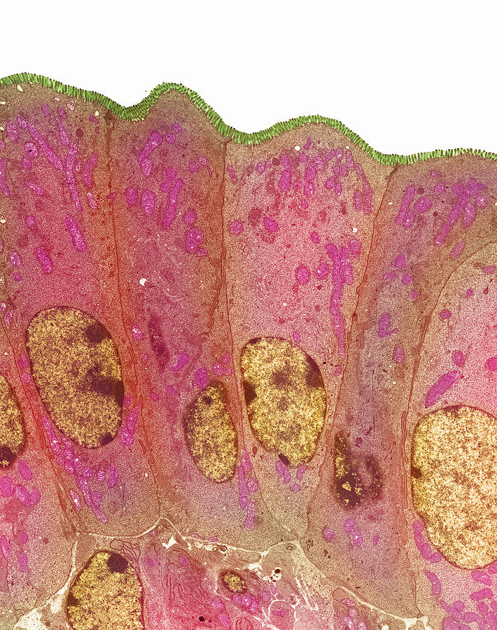
Duodenum secretory cells. Coloured transmission electron micrograph (TEM) of a section through the human duodenum, showing secretory cells of the... more
Title
Duodenum Secretory Cells #1
Artist
Steve Gschmeissner
Medium
Photograph
Description
Duodenum secretory cells. Coloured transmission electron micrograph (TEM) of a section through the human duodenum, showing secretory cells of the surface epithelium (lining). The duodenum is the first part of the small intestine. A row of columnar-shaped cells are seen, each with a rounded nucleus (brown) and mitochondria (pink) in the cytoplasm. Microvilli (green) appear as tiny projections from the surface of the cells (at top). Secretory cells secrete digestive enzymes, and an alkaline fluid into the pancreas which neutralises stomach acids. Microvilli serve to maximise the duodenum's surface area and hence its capacity to secrete. Magnification unknown.
Uploaded
May 6th, 2013
More from Steve Gschmeissner
Comments
There are no comments for Duodenum Secretory Cells #1. Click here to post the first comment.


