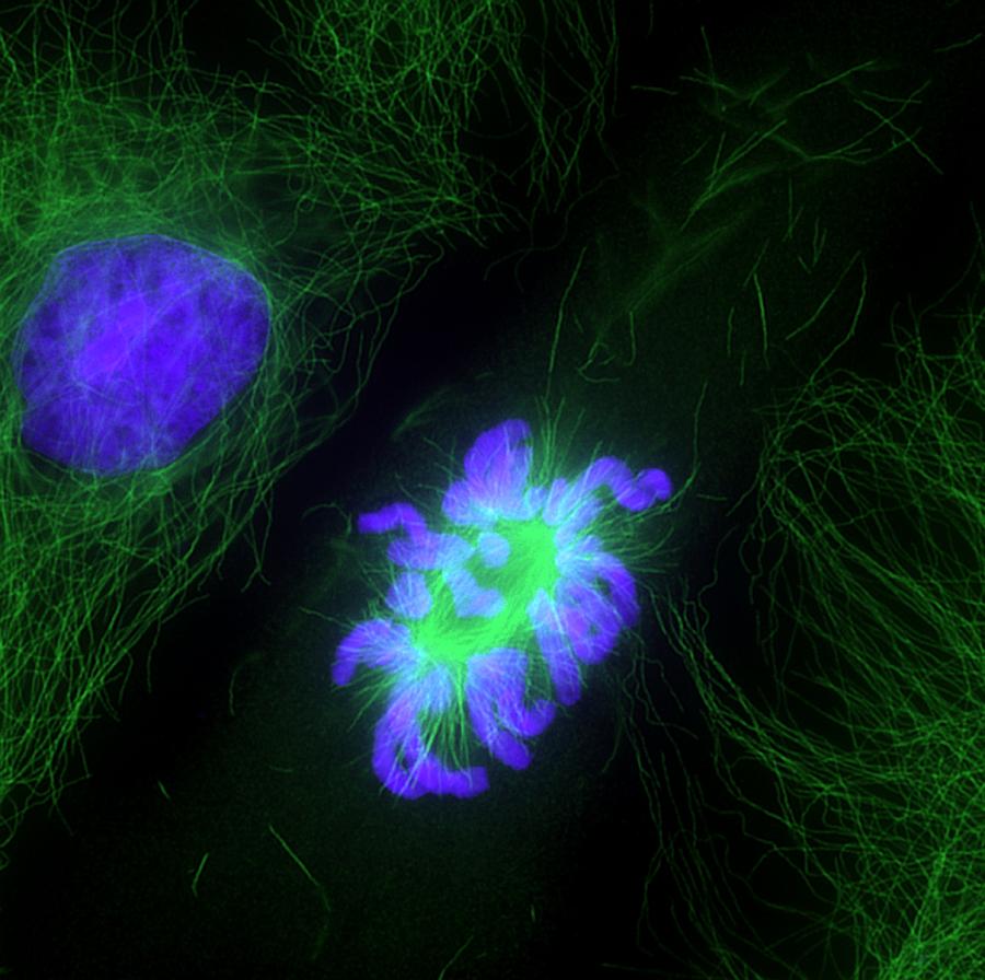
Cell Division, Fluorescent Micrograph is a photograph by Dr Torsten Wittmann which was uploaded on May 1st, 2013.
Cell Division, Fluorescent Micrograph
Cell division. Immunofluorescent light micrograph of a human epithelial cell (centre) during the prometaphase stage of mitosis. Mitosis is the cycle... more
Title
Cell Division, Fluorescent Micrograph
Artist
Dr Torsten Wittmann
Medium
Photograph
Description
Cell division. Immunofluorescent light micrograph of a human epithelial cell (centre) during the prometaphase stage of mitosis. Mitosis is the cycle of replication and division by which new body cells are formed. Here, the chromosomes of the parent cell (blue) have condensed, the nuclear envelope has broken down, and the microtubule cytoskeleton (green) has begun to reorganise itself in preparation for cytokinesis - when the chromosomes are pulled apart to opposite poles of the cell by the microtubules. When the cell has split, two identical daughter cells are formed.
Uploaded
May 1st, 2013
More from Dr Torsten Wittmann
Comments
There are no comments for Cell Division, Fluorescent Micrograph. Click here to post the first comment.


